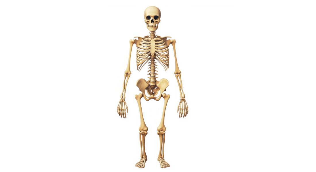
The Skeletal System
The skeletal system forms the framework of the human body, providing essential functions like support, protection, movement, storage, and blood cell formation.
Overview and Functions of the Skeletal System
The skeletal system has various functions:
- Support: Provides a framework for the body. For example, the legs support the body trunk, and the rib cage supports the chest wall.
- Protection: Protects delicate internal organs such as the brain (protected by the skull), heart and lungs (protected by the rib cage), and spinal cord (protected by the vertebral column).
- Movement: Muscles are attached to bones, which act as levers, enabling movement. However, bones cannot move on their own; they work with the muscular system.
- Storage:
- Fat Storage: Occurs in the yellow bone marrow of long bones.
- Mineral Storage: Bones store essential minerals like calcium and phosphorus, which can be released into the bloodstream when needed.
- Blood Cell Formation (Hematopoiesis): Occurs in the red bone marrow of long bones, producing red blood cells, white blood cells, and platelets.
Structure of the Human Skeleton
The adult human skeleton consists of 206 bones and is divided into:
- Axial Skeleton: The central core of the skeleton, including the skull, vertebral column, and rib cage. It provides support and protects the body's vital organs.
- Appendicular Skeleton: Comprises the bones of the upper and lower limbs, as well as the shoulder and pelvic girdles. These bones aid in movement.
Bone Tissue and Types of Bones
There are two main types of bone tissue:
- Compact Bone: Dense, smooth, and homogeneous in appearance. It forms the outer layer of bones and provides strength.
- Spongy Bone: Composed of small, needle-like pieces of bone with open spaces that resemble a sponge. It is lighter and provides some flexibility, especially at the ends of long bones.
In embryos, most of the skeleton is initially made of hyaline cartilage, which is gradually replaced by bone during development. However, cartilage remains in certain parts of the body, such as the nose, parts of the ribs, and joints.
Bone Growth and Remodeling
- Epiphyseal Plates: Located at the ends of long bones, these allow for bone growth during childhood. New cartilage forms continuously, and older cartilage is ossified (turned into bone), allowing bones to grow in length and width.
- Bone Remodeling: Involves two types of cells:
- Osteoblasts: "Bone builders" that form new bone tissue.
- Osteoclasts: "Bone destroyers" that break down bone tissue to remodel it and release stored calcium when needed.
Bone growth and remodeling are influenced by muscle activity. For example, bones get larger and thicker when large muscles are attached, but they become smaller and weaker with physical inactivity.
Classification of Bones
Bones are classified based on their shape:
- Long Bones: Longer than they are wide, consisting mostly of compact bone (e.g., femur, humerus).
- Short Bones: Cube-shaped, mostly spongy bone (e.g., wrist and ankle bones).
- Flat Bones: Thin, flat, and often curved, with two layers of compact bone and spongy bone in between (e.g., skull, ribs).
- Irregular Bones: Bones that don’t fit into the above categories (e.g., vertebrae, hip bones).
Anatomy of Long Bones
- Diaphysis: The shaft, made of compact bone.
- Epiphyses: The ends of long bones, composed of compact bone surrounding spongy bone. They are covered with articular cartilage, which reduces friction at joints.
- Periosteum: The outer covering of the bone shaft.
- Medullary Cavity: Found within the shaft, containing yellow marrow in adults (stores fat) and red marrow in infants (blood cell formation).
- Epiphyseal Line: The remnant of the epiphyseal plate, marking where growth occurred during development.
Bone Cells and Structure
- Osteon (Haversian System): A structural unit of compact bone consisting of concentric rings (lamellae) arranged around a central (Haversian) canal, which contains blood vessels and nerves.
- Lacunae: Small cavities within bone that house osteocytes (mature bone cells).
- Canaliculi: Tiny canals that radiate from the central canal to the lacunae, transporting nutrients to the osteocytes.
- Volkmann’s Canals: Perpendicular to the central canal, containing blood vessels and nerves, connecting different osteons.
Types of Joints and Movement
Joints (articulations) are classified based on their structure and function:
- Fibrous Joints: Immovable (e.g., sutures in the skull).
- Cartilaginous Joints: Can be immovable or slightly movable (e.g., the pubic symphysis and intervertebral discs).
- Synovial Joints: Freely movable and characterized by a joint cavity filled with synovial fluid. They are the most common type of joint in the body (e.g., knee, shoulder).
Synovial joints have associated structures:
- Bursae: Fluid-filled sacs that reduce friction between tissues.
- Tendon Sheaths: Elongated bursae that wrap around tendons experiencing significant friction.
Fractures and Bone Healing
Bone fractures are classified as:
- Simple (Closed) Fracture: The bone breaks cleanly but does not penetrate the skin.
- Compound (Open) Fracture: The broken bone penetrates the skin.
Bone healing occurs in several steps:
- Hematoma Formation: A blood-filled swelling forms at the fracture site.
- Fibrocartilage Callus Formation: Connective tissue forms a temporary splint.
- Bony Callus Formation: Osteoblasts and osteoclasts create a callus of spongy bone.
- Bone Remodeling: The bone is remodeled over time to return to its original shape.
Axial Skeleton
The axial skeleton consists of 80 bones and includes:
- Skull: Composed of 22 bones (frontal, parietal, temporal, etc.) that form the cranium and facial structures.
- Hyoid Bone: A single bone in the neck supporting the tongue.
- Vertebral Column: Composed of 32-34 vertebrae, which protect the spinal cord and support the body.
- Rib Cage: Comprising 24 ribs and the sternum, the rib cage protects the heart and lungs.
Appendicular Skeleton
The appendicular skeleton includes the bones of the limbs and girdles:
- Pectoral Girdle: Clavicle and scapula (shoulder girdle).
- Pelvic Girdle: The hip bones.
- Upper Limbs: Humerus, radius, ulna, carpals, metacarpals, and phalanges.
- Lower Limbs: Femur, tibia, fibula, tarsals, metatarsals, and phalanges.

Free Videos