The Digestive System
Overview of the Digestive System
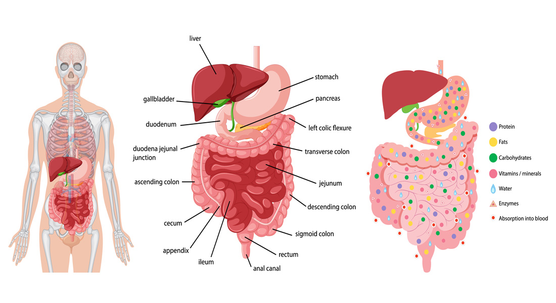
The digestive system, also known as the gastrointestinal (GI) tract or alimentary canal, is an organ system responsible for the ingestion, digestion, absorption of nutrients, and expulsion of waste. It involves a series of organs that work together to break down food, absorb nutrients, and eliminate waste products. The digestive system can be divided into the main digestive tract organs and accessory digestive organs.
Major Organs of the Digestive System
- Mouth
- Pharynx
- Esophagus
- Stomach
- Small Intestine
- Large Intestine
- Rectum
Accessory Digestive Organs
- Liver
- Gallbladder
- Pancreas
- Salivary Glands
Functions of the GI Tract
- Ingestion: The process of taking food into the alimentary canal, involving both eating and drinking.
- Propulsion: The movement of food through the GI tract, which includes both mixing and moving the contents along the alimentary canal.
- Digestion:
- Mechanical Digestion: The physical breakdown of food, such as chewing (mastication).
- Chemical Digestion: The breakdown of food into smaller molecules by enzymes.
- Absorption: The process by which digested food substances pass through the walls of certain organs in the alimentary canal into the bloodstream for distribution throughout the body.
- Elimination: The expulsion of indigestible food substances as feces through defecation.
Mouth
The mouth, also known as the oral cavity, is the initial portion of the alimentary canal that receives food and begins the digestive process by producing saliva.
Anatomy and Relations of the Mouth
- Anterior: Bounded by the lips.
- Posterior: Continues with the oropharynx.
- Lateral: Bounded by the muscles of the cheeks.
- Superior: Bounded by the bony hard palate.
- Inferior: Consists of the muscular tongue and soft tissues of the floor of the mouth.
Palate
- Hard Palate: The bony anterior portion forming the roof of the mouth.
- Soft Palate: The posterior portion, which is softer and consists of muscle and mucous membrane.
- Uvula: A small, curved fold of muscle covered with mucous membrane that hangs down from the middle of the soft palate.
Tongue
The tongue is a muscular organ in the mouth that plays a crucial role in manipulating food for mastication, swallowing, and is the primary organ of taste in the gustatory system.
Anatomy of the Tongue
- Dorsum (Upper Surface): Covered by taste buds housed in numerous lingual papillae.
- Divisions: The tongue is divided into two parts:
- Oral Part: Located at the front.
- Pharyngeal Part: Located at the back.
Blood Supply
- Arteries: The tongue receives blood from the lingual artery, which is a branch of the external carotid artery.
- Veins: Venous drainage is primarily through the lingual veins into the internal jugular vein.
Nerve Supply
- Motor Function: Hypoglossal nerve.
- Taste and Sensation: Glossopharyngeal nerve.
Functions of the Tongue
- Mastication (Chewing): The tongue helps to position food between the teeth.
- Deglutition (Swallowing): Assists in pushing food to the back of the mouth to initiate swallowing.
- Speech: Essential for articulation and pronunciation.
- Taste: Detects different flavors via taste buds.
Teeth
Human teeth are specialized structures in the mouth that function to mechanically break down food by cutting, tearing, crushing, and grinding, preparing it for swallowing and digestion.
Types of Teeth
- Incisors: Sharp, cutting teeth located at the front.
- Canines: Pointed teeth used for tearing food.
- Premolars: Teeth with flat surfaces for crushing food.
- Molars: Large, flat teeth at the back of the mouth used for grinding food.
Primary (Deciduous) Teeth
- Dental Formula: 2.1.0.2/2.1.0.2
- Number: 20 teeth (10 in the maxilla and 10 in the mandible).
- Eruption: Begins around 6 months of age.
- Types: Include central and lateral incisors, one canine, and two molars (first and second).
Permanent Teeth
- Dental Formula: 2.1.2.3/2.1.2.3
- Number: 32 teeth (16 in the maxilla and 16 in the mandible).
- Eruption: Usually completed by age 21.
- Types: Include two incisors (central and lateral), one canine, two premolars (first and second), and three molars (first, second, and third).
Parts of a Tooth
- Enamel: The hardest, most mineralized substance in the body, covering the crown of the tooth.
- Dentin: The layer beneath the enamel and cementum, forming the bulk of the tooth.
- Cementum: A specialized, bone-like substance covering the root of the tooth.
- Dental Pulp: The innermost part of the tooth, containing soft connective tissue, blood vessels, and nerves.
Functions of Teeth
- Incisors: Cutting food.
- Canines: Tearing food.
- Premolars: Crushing food.
- Molars: Grinding food.
Tooth Eruption
- Primary Teeth: Erupt between 6 months and 2 years of age.
- Permanent Teeth: Typically erupt between 6 years and 21 years of age.
Blood Supply, Venous Drainage, and Nerve Supply
- Blood Supply: Maxillary arteries supply blood to the relevant structures in the oral cavity and related areas.
- Venous Drainage: Blood from the oral cavity and associated structures is drained through the internal jugular veins.
- Nerve Supply: The maxillary nerves and mandibular nerves are responsible for the sensory and motor innervation of the oral structures.
Salivary Glands
The salivary glands are exocrine glands responsible for producing saliva, which is essential for digestion and maintaining oral hygiene. Humans have three pairs of major salivary glands and numerous minor salivary glands.
Major Salivary Glands
-
Parotid Glands:
- Location: Wrapped around the mandibular ramus.
- Function: Secretes saliva containing amylase to initiate the digestion of starches and facilitates mastication and swallowing.
- Duct: Saliva enters the oral cavity via the parotid duct.
- Significance: The largest of the salivary glands.
-
Submandibular Glands:
- Location: Beneath the lower jaws, superior to the digastric muscles.
- Function: Produces a mixture of serous fluid and mucus.
- Duct: Saliva enters the oral cavity via the submandibular duct.
-
Sublingual Glands:
- Location: Inferior to the tongue, anterior to the submandibular glands.
- Function: Primarily produces mucus; about 5% of saliva comes from these glands.
Minor Salivary Glands
- Location: Distributed throughout the oral cavity, particularly in the buccal, labial, and lingual mucosa.
- Number: Approximately 800 to 1,000 minor salivary glands.
Blood Supply and Venous Drainage
- Blood Supply: External carotid artery.
- Venous Drainage: Jugular veins.
Composition of Saliva
Daily saliva production amounts to approximately 1.5 liters. The composition includes:
- Water
- Mineral salts
- Enzyme (amylase)
- Mucus
- Lysozyme
- Immunoglobulins
Functions of Saliva
- Lubrication: Saliva coats the oral mucosa, protecting it from trauma during eating, swallowing, and speaking. Lack of saliva can lead to soreness and difficulty in food intake.
- Digestion: Saliva moistens food, helping to form a bolus that can be easily swallowed. It also contains enzymes that initiate the digestion process.
- Role in Taste: Acts as a medium for chemicals to reach taste receptors on the tongue.
- pH Maintenance: Saliva helps maintain the pH balance in the mouth, preventing tooth decay and other oral issues.
The Pharynx
The pharynx is the part of the throat situated behind the mouth and nasal cavity, and it plays a crucial role in swallowing by directing food from the mouth to the esophagus.
Function of the Pharynx
- Swallowing: Receives food from the mouth and directs it to the esophagus.
- Respiratory and Digestive Roles: Acts as a pathway for both air (to the lungs) and food (to the stomach).
The Esophagus
The esophagus is a muscular tube that connects the throat (pharynx) to the stomach, facilitating the movement of swallowed food.
Anatomy of the Esophagus
- Location: Runs behind the windpipe (trachea) and heart, in front of the spine.
- Dimensions: Approximately 25 cm in length and 2 cm in diameter.
Structure
- Layers: The esophagus consists of several layers:
- Mucosa: Stratified squamous epithelium, different from the single columnar layer of the stomach.
- Submucosa: Connective tissue.
- Muscle Fibers: Layers of muscle fibers between fibrous tissue, predominantly smooth muscle.
- Outer Layer: Connective tissue.
- Sphincters:
- Upper Esophageal Sphincter: Located at the top of the esophagus.
- Lower Esophageal Sphincter: Located at the junction with the stomach, preventing reflux of acidic stomach contents.
Functions of the Esophagus
- Formation of a Bolus: Ensures the food bolus is properly formed and directed toward the stomach.
- Swallowing: Facilitates the smooth passage of food from the pharynx to the stomach.
- Reducing Gastric Reflux: The constriction of the esophageal sphincters helps prevent the backflow of stomach acid into the esophagus.
Blood Supply and Venous Drainage
- Blood Supply: Esophageal arteries and inferior phrenic arteries.
- Venous Drainage: Left gastric vein.
Stomach
Anatomy and Location
- The stomach is a muscular organ located on the left side of the upper abdomen.
- It connects to the esophagus via the lower esophageal sphincter and to the small intestine via the pyloric sphincter.
- Relations:
- Anteriorly: Left lobe of the liver and anterior abdominal wall.
- Posteriorly: Abdominal aorta, pancreas, spleen, left kidney.
- Superiorly: Diaphragm, esophagus, left lobe of the liver.
- Inferiorly: Transverse colon, small intestine.
- Left Side: Diaphragm, spleen.
- Right Side: Liver, duodenum.
Structure
-
Regions: The stomach is divided into four regions:
- Cardiac Stomach (Cardia): Near the esophagus.
- Fundic Stomach (Fundus): Upper part, above the cardia.
- Body of Stomach: Main, central region.
- Pyloric Stomach (Pylorus): Near the small intestine.
-
Rugae: The inner lining of the stomach contains thick folds called rugae, which allow the stomach to expand and aid in digestion and absorption.
Functions
-
Digestion:
- Releases proteases (e.g., pepsin) and hydrochloric acid (HCl) to break down proteins and kill bacteria.
- The stomach churns food through peristalsis, a series of muscular contractions.
-
Absorption: Limited absorption occurs, primarily of small molecules such as water, alcohol, and certain medications.
Gastric Juice
- Composed of HCl, potassium chloride (KCl), and sodium chloride (NaCl).
- Provides an acidic environment (pH ~2) necessary for enzyme activity and protein digestion.
Gastric Secretory Cells
- Chief Cells: Secrete pepsinogen (inactive enzyme).
- Parietal Cells: Secrete HCl and intrinsic factor (necessary for vitamin B12 absorption).
- Mucous Cells: Secrete mucus and alkaline substances to neutralize stomach acid.
- G Cells: Secrete gastrin, a hormone that stimulates acid production.
Blood Supply and Venous Drainage
- Blood Supply: Right and left gastroepiploic arteries, gastric artery.
- Venous Drainage: Gastric vein.
Pancreas
Anatomy and Location
- The pancreas is a glandular organ located in the abdominal cavity, behind the stomach.
- It functions as both an exocrine (digestive) and endocrine (hormonal) gland.
- Length: Approximately 15 cm (6 inches).
Structure
- Divided into four parts:
- Head: Adjacent to the duodenum.
- Neck: Narrow section between the head and body.
- Body: The largest part, lies behind the pylorus.
- Tail: Extends towards the spleen.
Functions
- Endocrine Function: Regulation of blood glucose levels through hormone secretion:
- Alpha Cells: Secrete glucagon (raises blood glucose).
- Beta Cells: Secrete insulin (lowers blood glucose).
- Delta Cells: Secrete somatostatin (inhibits other pancreatic hormones).
- Gamma Cells: Secrete pancreatic polypeptide.
Blood Supply and Venous Drainage
- Blood Supply: Superior mesenteric artery, splenic artery.
- Venous Drainage: Superior mesenteric veins, splenic veins.
Liver
Anatomy and Location
- The liver is a large, reddish-brown organ located in the upper right quadrant of the abdomen.
- It plays a vital role in metabolism, detoxification, and digestion.
- Relations:
- Anteriorly: Diaphragm, anterior abdominal wall.
- Posteriorly: Esophagus, inferior vena cava, aorta, gall bladder, vertebral column, diaphragm.
- Laterally: Lower ribs, diaphragm.
- Superiorly: Diaphragm, anterior abdominal wall.
- Inferiorly: Stomach, bile ducts, duodenum, hepatic flexure of colon, right kidney.
Structure
- Weighs approximately 1.44–1.66 kg with a width of about 15 cm.
- Divided into right and left lobes by the falciform ligament.
Functions
- Metabolism: Carbohydrate, protein, and lipid metabolism.
- Synthesis: Produces proteins like albumin and clotting factors.
- Storage: Stores glycogen, vitamins A, D, B12, K, iron, and copper.
- Detoxification: Breaks down hormones and toxins.
- Bile Production: Produces bile, necessary for fat digestion.
Blood Supply and Venous Drainage
- Blood Supply: Hepatic artery.
- Venous Drainage: Hepatic veins.
Gallbladder
Anatomy and Location
- A small, pear-shaped organ located beneath the liver.
- Functions primarily as a storage site for bile.
Structure
- Length: 7-10 cm (2.8-3.9 inches).
- Diameter: 4 cm (1.6 inches).
- Capacity: Approximately 50 mL.
- Divided into three sections:
- Fundus: The broadest part.
- Body: Main storage area.
- Neck: Connects to the cystic duct.
Functions
- Stores and concentrates bile, releasing it into the small intestine during digestion.
Blood Supply and Venous Drainage
- Blood Supply: Cystic artery.
- Venous Drainage: Cystic veins.
Small Intestine
Anatomy and Structure
- Divided into three parts:
- Duodenum: C-shaped and approximately 20-25 cm in length.
- Jejunum: Middle section, about 2.5 m long.
- Ileum: Final section, about 3 m long with numerous villi.
Functions
- Digestion: The primary site for chemical digestion, aided by enzymes from the pancreas and liver.
- Absorption: The site where most nutrients from food are absorbed into the bloodstream.
- Immune Function: Supports the body’s immune system, particularly through gut flora.
Blood Supply and Venous Drainage
- Blood Supply: Celiac trunk, superior mesenteric artery.
- Venous Drainage: Superior mesenteric veins.
Large Intestine
Anatomy and Structure
- Also known as the colon, it is the final part of the digestive tract.
- Sections:
- Cecum: Beginning of the colon, connected to the ileum by the ileocecal valve.
- Ascending Colon: Travels upward.
- Transverse Colon: Crosses the abdominal cavity.
- Descending Colon: Travels downward.
- Sigmoid Colon: S-shaped and connects to the rectum.
- Rectum: Final section, stores feces.
Functions
- Absorption: Absorbs water and electrolytes from indigestible food matter.
- Gut Flora: Houses over 700 species of bacteria that aid in digestion and vitamin production.
- Storage and Elimination: Stores fecal matter until defecation.
Blood Supply and Venous Drainage
- Blood Supply: Superior mesenteric artery, inferior mesenteric artery.
- Venous Drainage: Superior mesenteric veins, inferior mesenteric veins.

Free Videos