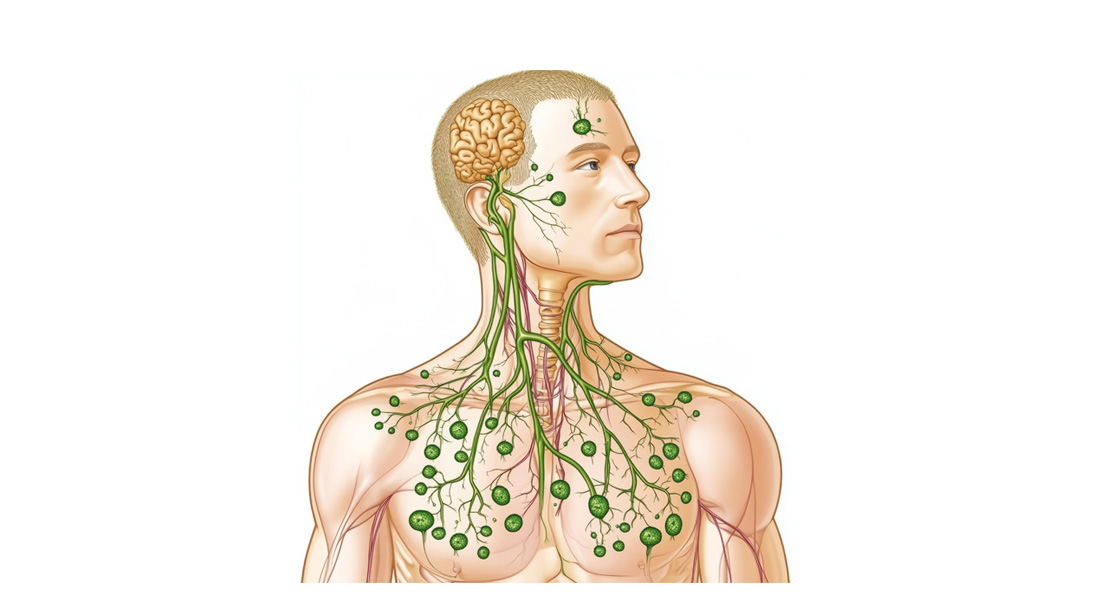
The Lymphatic System
The lymphatic system is a crucial network that contributes to maintaining fluid balance, defending the body against infections, and ensuring the proper functioning of the immune system. It consists of lymphatic vessels, lymph nodes, and various lymphoid organs and tissues.
Functions of the Lymphatic System
- Maintaining Fluid Balance: The lymphatic system returns excess fluid (approximately 3 liters/day) from tissues back to the blood circulation, ensuring blood volume remains stable.
- Immunity: It plays an essential role in immune defense by filtering out harmful substances and pathogens, enabling the body to fight infections and diseases.
Components of the Lymphatic System
-
Lymph:
- Definition: Lymph is a clear, watery fluid that forms when interstitial fluid (fluid in the spaces between cells) enters lymphatic vessels.
- Formation: As blood circulates, nutrients and gases are filtered from the plasma through capillary walls to form interstitial fluid. The excess fluid is drained into lymphatic vessels, where it becomes lymph.
- Composition: Lymph is compositionally similar to interstitial fluid, containing water, proteins, and waste products. In areas such as the small intestine, lymph also contains absorbed dietary fats, giving it a milky appearance (referred to as chyle).
-
Lymphatic Vessels:
- Structure: Lymph vessels are thin-walled, valved structures that carry lymph from tissues toward the heart. They begin as lymphatic capillaries, which merge to form larger vessels.
- Lymphatic Trunks: These vessels unite to form lymphatic trunks, which eventually drain into one of two ducts:
- Right Lymphatic Duct: Collects lymph from the upper right quadrant of the body.
- Thoracic Duct: The largest lymphatic vessel, which collects lymph from the rest of the body.
- Function: Lymphatic vessels return excess tissue fluid to the bloodstream and play a role in absorbing dietary fats from the small intestine through specialized vessels called lacteals.
-
Lymphatic Ducts:
- Thoracic Duct: Also known as the left lymphatic duct, it is the main channel for returning lymph to the bloodstream. It begins at an enlarged sac called the cisterna chyli (located at the anterior of the second lumbar vertebra) and drains lymph into the venous system at the junction of the left subclavian and internal jugular veins.
- Right Lymphatic Duct: This much smaller duct collects lymph from the right side of the head, neck, chest, and right arm, and empties it into the bloodstream at the junction of the right subclavian and internal jugular veins.
-
Lymph Nodes:
- Structure: Lymph nodes are small, bean-shaped organs that act as filters, trapping pathogens and debris as lymph passes through them. Each node is covered by a capsule and contains immune cells, including B cells and T cells, which actively respond to foreign invaders.
- Location: There are about 600 lymph nodes in the body, commonly found in clusters in regions like the cervical, axillary, and inguinal areas.
- Function: They filter lymph before it is returned to the bloodstream and are critical sites for mounting immune responses.
Structure of Lymphatic Vessels and Nodes
Lymphatic Capillaries:
- Structure: These are single-layered endothelial cells forming flap-like mini-valves that prevent the backward flow of lymph. The permeability of lymphatic capillaries is greater than that of blood capillaries, allowing large molecules, including proteins and fats, to enter.
Lymphatic Vessels:
- Layers: Like veins, lymphatic vessels have three layers:
- Tunica intima: The inner layer lined by endothelial cells.
- Tunica media: The middle layer composed of smooth muscle and elastic fibers, allowing the vessel to contract and propel lymph.
- Tunica adventitia: The outermost layer made of fibrous connective tissue.
- Valves: Lymphatic vessels contain valves that ensure one-way flow towards the heart.
Lymph Nodes:
- Capsule and Trabeculae: The outer capsule of the lymph node extends inward as trabeculae, dividing the node into compartments. The internal structure of the node is supported by a network of reticular fibers.
- Cortex and Medulla: The lymph node is divided into two regions:
- Cortex: Contains densely packed lymphocytes (mainly B cells).
- Medulla: Contains cords of lymphatic tissue, macrophages, and plasma cells.
- Afferent and Efferent Vessels: Lymph enters the node via afferent lymphatic vessels and exits through efferent vessels.
Lymphatic Trunks and Ducts
Lymphatic vessels drain into lymphatic trunks that further converge into one of two large ducts.
Major Lymphatic Trunks:
- Lumbar Trunks: Drain lymph from the lower limbs and pelvic region.
- Intestinal Trunk: Collects lymph from the digestive organs.
- Bronchomediastinal Trunks: Drain lymph from the thorax, lungs, and heart.
- Subclavian Trunks: Drain the upper limbs.
- Jugular Trunks: Drain the head and neck.
Lymphatic Ducts:
- Thoracic Duct: The largest lymphatic vessel that drains the left side of the body and the lower right side.
- Right Lymphatic Duct: Drains the right upper quadrant of the body.
Specialized Structures and Functions
- Lacteals: Specialized lymphatic capillaries in the small intestine that absorb dietary fats and transport them to the bloodstream. The lymph within these capillaries is milky white due to the presence of fats and is called chyle.
- Cisterna Chyli: An enlarged sac that collects lymph from the lower limbs and digestive organs before transporting it to the thoracic duct.
Deep Regional Cervical Lymph Nodes
The deep regional cervical lymph nodes form an inner circle surrounding the larynx, trachea, and pharynx. This group includes:
-
Prelaryngeal and Pretracheal Nodes
- Prelaryngeal nodes are situated on the cricothyroid membrane.
- Pretracheal nodes lie in front of the trachea, below the isthmus of the thyroid gland.
- These nodes drain the larynx, trachea, and the isthmus of the thyroid, as well as lymph from the anterior cervical nodes of the superficial regional cervical lymph nodes. They drain into nearby deep cervical nodes.
-
Paratracheal Nodes
- Located on the sides of the trachea and esophagus.
- They drain lymph from the esophagus, trachea, and larynx and also into deep cervical nodes.
-
Retropharyngeal Nodes
- Positioned in front of the prevertebral fascia and behind the buccopharyngeal fascia, covering the posterior wall of the pharynx.
- These nodes drain from the pharynx, auditory tube, soft palate, posterior part of the hard palate, and nose, before draining into the jugulodigastric node.
Axillary Lymph Nodes
Located in the armpit region, the axillary lymph nodes are divided into superficial and deep groups. These nodes are responsible for draining lymph from the arms, chest wall, and breast. They are classified into five main groups:
- Pectoral (anterior) group: Found along the lower border of the pectoralis minor muscle, draining from the upper half of the anterior chest wall and the major part of the breast.
- Scapular (posterior) group: Situated along the posterior fold of the axilla, draining from the posterior wall of the trunk and the axillary tail of the breast.
- Lateral group: Located medial to the axillary vein, draining lymph from the upper limb.
- Central group: Found in the upper axilla, receiving lymph from the pectoral, scapular, and lateral groups.
- Apical or infraclavicular group: Positioned along the axillary vessel below the clavicle, draining lymph from the central group and the upper part of the breast.
Thoracic Lymph Nodes
Thoracic lymph nodes are categorized into parietal and visceral lymph nodes:
-
Parietal Lymph Nodes
- Include parasternal, intercostal, and diaphragmatic nodes.
- Parasternal nodes are located along the sides of the internal mammary artery, draining the mammary glands, anterior abdominal wall, and upper surface of the liver.
- Intercostal nodes are found in the posterior part of intercostal spaces, draining the posterior and lateral parts of the chest wall.
- Diaphragmatic nodes are located on the upper surface of the diaphragm, divided into anterior, middle, and posterior sets, receiving lymph from the diaphragm, pericardium, pleura, and upper liver surface.
-
Visceral Lymph Nodes
- Include tracheobronchial and mediastinal nodes.
- Tracheobronchial nodes are found around the trachea and bronchi, consisting of pulmonary, bronchopulmonary, and tracheobronchial nodes.
- Pulmonary lymph nodes lie within the lungs, draining lymph from the lungs and emptying into the bronchopulmonary nodes.
- Mediastinal nodes are divided into anterior and posterior groups, helping in the production of mature lymphocytes from the bone marrow and thymus.
Other Lymph Node Groups
-
Supratrochlear Nodes (Cubital Lymph Nodes): Located near the elbow and the medial epicondyle of the humerus, these nodes drain from the fingers, ulnar side of the arm, and superficial forearm. They drain into the axillary nodes.
-
Inguinal Lymph Nodes: These nodes are found in the groin and may be either superficial or deep. They drain from the genital areas, buttocks, anus, abdominal wall, and legs.
-
Femoral Nodes: Located in the upper thigh, along the femoral vein, they drain parts of the genitals, buttocks, thigh, and medial leg.
-
Popliteal Nodes: Found in the popliteal fossa (behind the knee), they drain lymph from the knee, thigh, calf, and feet.
Lymphoid Organs
The lymphoid organs are critical structures in the body’s immune system, playing a key role in protecting the body from pathogens and maintaining immune function. These include the spleen, thymus, tonsils, and Peyer’s patches.
Spleen
- Largest lymphoid organ, located in the left hypochondrium of the abdominal cavity.
- Oval-shaped, purplish, soft, and highly vascular, about 12 cm long and weighing around 200 grams.
- Structure:
- Fibrous capsule encloses the spleen, and inward extensions of this capsule, called trabeculae, divide the spleen into compartments.
- Parenchyma contains two types of tissue:
- White pulp (immune function, contains lymphocytes and macrophages around central arteries)
- Red pulp (involved in filtering blood, contains splenic sinusoids and splenic cords rich in RBCs, macrophages, and immune cells).
- Destruction of old or damaged RBCs and blood-borne pathogens occurs in the red pulp.
- Blood Supply: Arterial supply is through the splenic artery, and venous drainage occurs via the splenic vein.
- Lymphatic Drainage: Few lymphatics arise from the capsule and trabeculae, draining into pancreaticosplenic lymph nodes.
Thymus
- Location: In the mediastinum, between the sternum and the aorta.
- Structure:
- Reddish, lobed structure, weighing about 30-40 g at puberty; it starts to atrophy after puberty, weighing about 3 g in old age.
- Each lobe is divided into lobules by fibrous extensions (trabeculae), and each lobule contains an outer cortex (immature T cells, macrophages) and an inner medulla (fewer mature T cells, thymic corpuscles).
- Thymic corpuscles are structures composed of concentric clusters of keratinized epithelial cells.
- Function: The thymus is the site of T cell maturation. Immature T cells (thymocytes) undergo selection and differentiation here before being released into circulation.
Tonsils
- Tonsils form a ring of lymphoid tissue around the entrance to the pharynx and include:
- Pharyngeal tonsil (adenoids), located in the nasopharynx.
- Palatine tonsils, located at the posterior region of the oral cavity.
- Lingual tonsils, at the base of the tongue.
- Tubal tonsils, near the openings of the auditory tubes.
- Function: Tonsils trap and destroy pathogens entering through food or air. They contain lymphoid follicles and tonsillar crypts, which increase surface area to enhance pathogen trapping.
Peyer’s Patches
- Found in the walls of the small intestine, especially in the ileum.
- Structurally similar to tonsils, consisting of large clusters of lymphoid follicles.
- Function: Peyer’s patches monitor intestinal bacteria populations and prevent the growth of pathogenic bacteria, thereby contributing to immune defense in the digestive system.

Free Videos