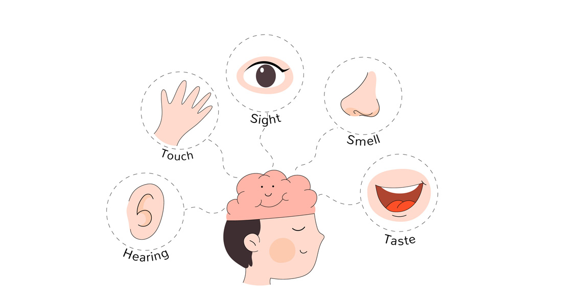
The Sensory Organs
The sensory organs in humans, focusing on the five primary senses: sight, hearing, smell, taste, and touch.
1. The Eye (Sense of Sight)
The eye is the sensory organ for vision, allowing us to detect light, colors, and shapes.
Structure of the Eye:
- Sclera: The tough, white outer covering of the eye that protects it.
- Cornea: The clear, dome-shaped surface that covers the front of the eye, helping to focus light.
- Iris: The colored part of the eye, controlling the amount of light that enters by adjusting the size of the pupil.
- Pupil: The black circular opening in the center of the iris, allowing light to enter the eye.
- Lens: A transparent structure behind the pupil that changes shape to focus light onto the retina.
- Retina: The innermost layer of the eye, containing photoreceptors:
- Rods: Detect dim light and are crucial for night vision.
- Cones: Responsible for color vision and sharpness.
Mechanism of Vision:
- Light enters through the cornea and is refracted (bent).
- The iris adjusts the size of the pupil to control light entry.
- The lens further focuses the light, directing it onto the retina.
- Photoreceptor cells in the retina convert the light into electrical impulses.
- These impulses are sent to the brain's occipital lobe via the optic nerve for interpretation.
Eyelids and Eyelashes
- Eyelids are movable folds of skin composed of skeletal muscle (orbicularis oculi), situated above and below the front of each eye. Their primary function is to protect the eyes from dust and debris.
- The free edges of the eyelids contain eyelashes, short curved hairs that help prevent foreign particles from entering the eye.
- Conjunctiva is a thin, transparent membrane that lines the inside of the eyelids and covers the sclera (white of the eye), protecting the cornea. It is composed of columnar epithelium, contributing to the protection and lubrication of the eye.
- Blinking spreads tears over the eye’s surface, keeping it moist and removing irritants, while the eyelashes further act as a defense mechanism by trapping dust and debris.
Eyebrows
- Eyebrows play an essential role in shading the eyes from sunlight and diverting sweat away from the eyes. This prevents irritation and maintains clear vision.
Lacrimal Apparatus
- The lacrimal apparatus consists of the lacrimal glands, which are almond-shaped and located in the upper outer region of each eye. They produce tears that lubricate, clean, and protect the surface of the eye.
- Lacrimal ducts transport tears to the anterior surface of the eyeball, and blinking helps spread these tears across the eye.
- Tears consist of water, mucus, sodium chloride (around 1%), mineral salts, lysozyme (a bactericidal enzyme), and gamma globulin. These components allow tears to protect, lubricate, and nourish the eye.
- Functions of Tears: Regular secretion of tears (around 1 mL daily) is important for cleaning, moistening, and providing nutrients to the eyeball. Increased tear production occurs in response to irritants, such as chemicals or emotional stimuli.
Layers of the Eye
The eye is composed of three layers:
1. Fibrous Layer (Outer Layer)
-
Sclera: The white, fibrous outermost layer of the eye that provides structure and protection. It is composed of tough connective tissue.
-
Cornea: The transparent front part of the sclera that allows light to enter the eye. It plays a crucial role in focusing light onto the retina.
2. Vascular Layer (Middle Layer)
-
Choroid: Contains blood vessels and melanin pigment. The melanin absorbs excess light, preventing reflection and scattering within the eye, thus maintaining image clarity.
-
Ciliary Body: A ring-shaped structure that produces aqueous humor and contains muscles responsible for changing the shape of the lens to focus light.
-
Iris: The colored part of the eye that controls the diameter of the pupil (the opening in the center of the iris), regulating the amount of light that enters the eye.
3. Nervous Layer (Inner Layer)
-
Retina: Contains the photoreceptor cells (rods and cones) that detect light and color.
-
Rods: Approximately 120 million cells, located toward the periphery of the retina, responsible for vision in low-light conditions and detecting black and white.
-
Cones: About 6-7 million cells located mainly in the central retina (macula lutea), responsible for detecting color (red, green, and blue) and providing sharp, detailed vision.
-
-
Fovea Centralis: A small depression in the macula lutea where vision is the sharpest. This is the point where light is focused when looking directly at an object.
-
Optic Disk (Blind Spot): The area where the optic nerve leaves the eye, containing no photoreceptors.
Lens and Its Role in Focusing
- The lens is a biconvex, transparent structure made of elastic proteins, located behind the pupil. It focuses light onto the retina by changing shape through the action of the ciliary muscle.
- For near objects, the ciliary muscle contracts, increasing the lens's curvature, thereby increasing its refractive power (thickness).
- For distant objects, the ciliary muscle relaxes, flattening the lens and reducing its refractive power.
Pupil and Light Regulation
- The pupil is the opening in the center of the iris that controls how much light enters the eye. The size of the pupil is regulated by two types of muscles:
- Circular muscles: Contract in response to bright light, reducing the pupil size (pupil constriction).
- Radial muscles: Contract in dim light, enlarging the pupil (pupil dilation).
- This process, controlled by the autonomic nervous system, ensures that the right amount of light reaches the retina for optimal vision.
Visual Pathway
- Light entering the eye is detected by the photoreceptors (rods and cones) in the retina.
- These photoreceptors synapse with bipolar cells, which in turn synapse with ganglion cells.
- The axons of the ganglion cells form the optic nerve, which transmits visual information to the thalamus and then to the visual cortex in the occipital lobe of the brain, where the information is processed.
Compartments of the Eye
- The eyeball is divided into two main compartments:
- Anterior Chamber: Located between the cornea and the lens, filled with aqueous humor produced by the ciliary body. This fluid maintains intraocular pressure and nourishes the eye.
- Posterior Chamber: Located behind the lens, filled with a gel-like substance called vitreous humor that supports the structure of the eye and helps maintain its shape.
Both the aqueous and vitreous humors also contribute to the refractive properties of the eye, helping focus light onto the retina.
Mechanism of Vision
-
Light Refraction in the Eye:
- Refraction refers to the bending of light as it passes through different mediums. In the eye, light is refracted through several structures, including the cornea, aqueous humor, lens, and vitreous humor. This bending of light focuses it onto the retina for clear vision.
- The lens is the only adjustable part of this system, capable of changing its shape to focus light on near or distant objects. This process, known as accommodation, is vital for maintaining focus as the distance of objects changes.
-
Retinal Response:
- When light strikes the retina, photoreceptor cells (rods and cones) trigger chemical reactions:
- Rods (responsible for vision in dim light) contain the pigment rhodopsin, which breaks down into scotopsin and trans-retinal upon light absorption.
- Cones (responsible for color vision) contain iodopsin, which breaks down into photopsin and trans-retinal upon absorbing different wavelengths of light.
- These reactions generate nerve impulses that are transmitted to the bipolar cells, then to ganglion cells. The axons of the ganglion cells form the optic nerve, which carries the information to the brain.
- When light strikes the retina, photoreceptor cells (rods and cones) trigger chemical reactions:
-
Image Processing:
- The image formed on the retina is inverted (upside down). The optic nerves from both eyes meet at the optic chiasma, where fibers cross, allowing each visual area in the brain to receive input from both eyes.
- This crossing enables binocular vision, which integrates depth and three-dimensional perception. The brain processes the inverted images and flips them right side up, allowing us to perceive the world accurately.
Common Visual Disorders
-
Conjunctivitis:
- Also known as pink eye, this is an inflammation of the conjunctiva, the membrane covering the front of the eye and lining the eyelids. It can be caused by bacterial or viral infections, allergies, or irritants, leading to red, itchy, and watery eyes.
- Also known as pink eye, this is an inflammation of the conjunctiva, the membrane covering the front of the eye and lining the eyelids. It can be caused by bacterial or viral infections, allergies, or irritants, leading to red, itchy, and watery eyes.
-
Hypermetropia (Farsightedness):
- In this condition, distant objects can be seen clearly, but near objects are blurry. The eye focuses light behind the retina due to a flattened lens. Correction involves convex lenses to properly focus light on the retina.
- In this condition, distant objects can be seen clearly, but near objects are blurry. The eye focuses light behind the retina due to a flattened lens. Correction involves convex lenses to properly focus light on the retina.
-
Myopia (Nearsightedness):
- Individuals with myopia can see near objects well, but distant objects appear blurry. The eye focuses light in front of the retina, often due to an elongated eyeball or thickened lens. Concave lenses are used to correct this condition.
- Individuals with myopia can see near objects well, but distant objects appear blurry. The eye focuses light in front of the retina, often due to an elongated eyeball or thickened lens. Concave lenses are used to correct this condition.
-
Presbyopia:
- A result of aging, presbyopia is the inability to focus on near objects due to the loss of elasticity in the lens. The lens becomes less able to change shape, making reading or close work difficult. Reading glasses or bifocals are often required.
- A result of aging, presbyopia is the inability to focus on near objects due to the loss of elasticity in the lens. The lens becomes less able to change shape, making reading or close work difficult. Reading glasses or bifocals are often required.
-
Astigmatism:
- Astigmatism occurs when the cornea or lens has an irregular curvature, causing light rays to scatter rather than focus on a single point on the retina. This results in blurred or distorted vision. Specially curved lenses are used to correct the condition.
- Astigmatism occurs when the cornea or lens has an irregular curvature, causing light rays to scatter rather than focus on a single point on the retina. This results in blurred or distorted vision. Specially curved lenses are used to correct the condition.
-
Glaucoma:
- A group of disorders characterized by increased intraocular pressure that damages the optic nerve and can lead to vision loss. Elevated pressure in the anterior cavity affects the vitreous humor, which transmits pressure to the retina and optic nerve. Glaucoma can cause halos around lights and loss of peripheral vision. Risk factors include high blood pressure and diabetes.
- A group of disorders characterized by increased intraocular pressure that damages the optic nerve and can lead to vision loss. Elevated pressure in the anterior cavity affects the vitreous humor, which transmits pressure to the retina and optic nerve. Glaucoma can cause halos around lights and loss of peripheral vision. Risk factors include high blood pressure and diabetes.
-
Night Blindness:
- Also known as nyctalopia, night blindness is the inability to see in low light conditions, often caused by a deficiency in rod function. Common causes include vitamin A deficiency, glaucoma, and cataracts.
- Also known as nyctalopia, night blindness is the inability to see in low light conditions, often caused by a deficiency in rod function. Common causes include vitamin A deficiency, glaucoma, and cataracts.
-
Cataract:
- A cataract is the clouding of the lens, leading to decreased vision. It is commonly associated with aging but can also result from trauma, radiation exposure, or certain diseases. Symptoms include blurred vision, sensitivity to light, and difficulty seeing at night. Cataracts are usually treated by surgically replacing the lens with an artificial one.
2. The Ear (Sense of Hearing and Balance)
The ear is responsible for detecting sound and maintaining balance.
Structure of the Ear:
- Outer Ear:
- Pinna: The visible part of the ear that collects sound waves.
- Auditory Canal: Channels sound waves to the eardrum.
- Middle Ear:
- Tympanic Membrane (Eardrum): Vibrates when sound waves hit it.
- Ossicles: Three small bones (malleus, incus, and stapes) that amplify sound vibrations.
- Inner Ear:
- Cochlea: A spiral-shaped structure filled with fluid and lined with tiny hair cells that convert sound vibrations into electrical signals.
- Vestibular Apparatus: Includes the semicircular canals and otolith organs (utricle and saccule), which help maintain balance by detecting head position and movement.
Mechanism of Hearing:
- Sound waves are collected by the pinna and travel down the auditory canal to the eardrum.
- The eardrum vibrates, transmitting sound to the ossicles.
- The ossicles amplify the sound and pass it to the cochlea.
- In the cochlea, fluid moves and bends the hair cells, creating electrical signals.
- These signals travel to the auditory cortex in the brain via the auditory nerve, where they are interpreted as sound.
Mechanism of Balance:
- The vestibular system detects head movements and changes in position.
- Fluid movement in the semicircular canals and otolith organs stimulates hair cells.
- The brain receives these signals, helping to maintain equilibrium.
The Anatomy and Functions of the Ear
1. Outer Ear:
The outer ear consists of the auricle (pinna) and the ear canal:
- Auricle (Pinna): Made of elastic cartilage, it functions as a funnel, capturing sound waves and directing them toward the external auditory canal.
- External Auditory Canal: Lined with small hairs and ceruminous glands that secrete earwax (cerumen), which serves as a protective barrier, trapping dust and foreign particles to prevent them from reaching the delicate eardrum. This canal leads the sound waves to the tympanic membrane (eardrum).
2. Middle Ear:
The middle ear is an air-filled cavity that contains three small bones known as the auditory ossicles and the auditory tube:
-
Auditory Ossicles:
- Malleus: Hammer-shaped, it connects to the eardrum and transmits vibrations to the incus.
- Incus: Anvil-shaped, it passes vibrations from the malleus to the stapes.
- Stapes: Stirrup-shaped, it connects with the oval window of the inner ear, transmitting vibrations.
These bones amplify sound vibrations, allowing effective transmission from the outer ear to the inner ear.
-
Auditory Tube (Eustachian Tube): This tube connects the middle ear to the pharynx (throat). Its function is to equalize air pressure between the middle ear and the external environment, ensuring that pressure changes do not interfere with hearing.
3. Inner Ear:
The inner ear consists of the cochlea, vestibule, and semicircular canals:
-
Cochlea: A spiral-shaped organ responsible for hearing. It converts sound vibrations into nerve impulses. It contains three compartments: the scala vestibuli (with perilymph), the cochlear duct (with endolymph), and the scala tympani (with perilymph).
Within the cochlear duct lies the basilar membrane, where hair cells act as auditory receptors. These hair cells detect sound vibrations and generate electrical impulses transmitted via the vestibulocochlear nerve to the auditory areas of the brain.
-
Vestibule and Semicircular Canals: These structures are responsible for balance and equilibrium. The vestibule detects changes in head position, while the semicircular canals detect rotational movement, aiding in maintaining body posture.
Hearing Process:
The process of hearing involves the following steps:
- Sound waves enter the auricle and pass through the external auditory canal.
- The waves strike the tympanic membrane, causing it to vibrate.
- These vibrations are transmitted through the malleus, incus, and stapes to the oval window.
- The oval window creates pressure waves in the perilymph within the scala vestibuli.
- These pressure waves travel through the cochlea, vibrating the basilar membrane and stimulating hair cells.
- Hair cells generate nerve impulses, which travel through the vestibulocochlear nerve to the brain, where the auditory cortex interprets them as sound.
Common Disorders of the Ear:
-
Motion Sickness: Characterized by nausea and dizziness, it occurs when the brain receives conflicting signals from the inner ear and the visual system about motion and balance.
-
Deafness: This can result from damage to the eardrum, inner ear hair cells, or auditory nerve. Causes may include infections, loud noises, or age-related deterioration.
-
Otitis Media: A middle ear infection common in children, often accompanied by fever and hearing loss due to fluid buildup behind the eardrum.
-
Hypermetropia (Far-sightedness): A condition where the patient has difficulty seeing near objects but can see distant ones clearly.
-
Myopia (Near-sightedness): Opposite of hypermetropia, in this condition, the patient has trouble seeing distant objects.
-
Presbyopia: Age-related loss of elasticity in the lens, causing difficulty in focusing on close objects.
-
Astigmatism: A defect in the curvature of the cornea or lens, leading to blurry or distorted vision.
-
Glaucoma: A group of eye conditions that damage the optic nerve, often due to high intraocular pressure.
-
Night Blindness: Difficulty seeing in low-light conditions, often due to vitamin A deficiency or retinal disorders.
-
Cataract: A clouding of the lens, leading to blurred vision and light sensitivity.
3. The Nose (Sense of Smell)
The nose is the organ for detecting airborne chemicals, contributing to the sense of smell.
Structure of the Nose:
- Nasal Cavity: The hollow space behind the nose where air enters.
- Olfactory Epithelium: A specialized tissue at the roof of the nasal cavity that contains olfactory receptor cells.
- Olfactory Bulb: A structure located just above the nasal cavity that processes smell information.
Mechanism of Smell:
- Odor molecules enter the nasal cavity and dissolve in the mucus lining.
- These molecules bind to olfactory receptors in the olfactory epithelium.
- The receptors send electrical signals to the olfactory bulb.
- The signals are relayed to the olfactory cortex in the brain, where smells are identified.
The Anatomy of the Nose
The nose is the primary organ responsible for the sense of smell and plays a significant role in respiration. It can be divided into external nose and nasal cavity. Together, these structures perform important respiratory and olfactory functions.
External Nose:
-
Structure: The external nose consists of bone, cartilage, and skin. Its main components include:
- Nasal bones: These form the upper part of the nose.
- Nasal cartilage: This makes up the lower, flexible part of the nose, allowing for movement and flexibility.
- Nostrils (nares): Openings that lead to the nasal cavity, surrounded by the alae (the flared portion on each side).
- Septum: The midline structure made of cartilage and bone that separates the nostrils.
-
Function: The external nose acts as a passage for air to enter and exit the body, and also contains sensory receptors responsible for the sense of smell.
Nasal Cavity:
The nasal cavity is the internal chamber of the nose and is divided into two halves by the nasal septum. It serves as a pathway for air and helps condition the air by filtering, warming, and moistening it before it enters the lungs.
-
Boundaries:
- Roof: Formed by parts of the frontal, ethmoid, and sphenoid bones.
- Floor: Composed of the hard palate (made of maxillary and palatine bones).
- Medial Wall: The nasal septum divides the nasal cavity into two chambers.
- Lateral Wall: Contains three prominent bony projections called conchae (superior, middle, and inferior), which increase surface area and help with air filtration.
-
Nasal Conchae (Turbinates):
- Superior, middle, and inferior conchae are curved bony structures that project into the nasal cavity from the lateral walls. They increase the surface area within the nasal cavity, allowing more efficient conditioning of the air.
- The conchae divide the nasal cavity into air passages called meatuses (superior, middle, and inferior meatuses), which direct airflow and improve the exposure of air to the mucous membrane.
-
Nasal Mucosa:
- The nasal cavity is lined with respiratory epithelium, which is rich in goblet cells that secrete mucus.
- Cilia on the epithelial cells help move mucus and trapped particles out of the nasal passages, keeping the airway clear.
- The mucous membrane also has a dense network of blood vessels, helping to warm and moisten incoming air.
-
Olfactory Epithelium:
- Located in the upper part of the nasal cavity, the olfactory epithelium contains specialized receptors for the sense of smell.
- These olfactory receptors send signals to the olfactory bulb, which then transmits sensory information to the brain for processing smells.
-
Nasal Septum:
- The septum divides the nasal cavity into two halves. It is made of cartilage in the front and bone in the back.
- It plays an important role in supporting the structure of the nose and directing airflow.
Paranasal Sinuses:
The nose is connected to four pairs of paranasal sinuses, which are air-filled cavities within the facial bones. They are named based on the bones in which they are located:
- Frontal Sinus (located in the forehead)
- Ethmoid Sinus (between the eyes)
- Sphenoid Sinus (deep behind the nose)
- Maxillary Sinus (located in the cheekbones)
- Function: These sinuses help lighten the weight of the skull, produce mucus, and contribute to the resonance of voice. They also drain into the nasal cavity through small openings.
Blood Supply:
The nose has a rich blood supply derived from branches of both the internal carotid artery and external carotid artery. Some important vessels include:
- Sphenopalatine artery
- Ethmoidal arteries
- Facial artery
The high density of blood vessels in the nasal cavity aids in warming the air. However, it also makes the nose prone to nosebleeds (epistaxis), particularly in the Kiesselbach's area (an area rich in blood vessels located at the front of the nasal septum).
Nerve Supply:
- Olfactory Nerve (Cranial Nerve I): Responsible for transmitting the sense of smell from the nasal cavity to the brain.
- Trigeminal Nerve (Cranial Nerve V): Provides sensory innervation to the nasal cavity, including pain and temperature sensations.
- Ophthalmic and Maxillary divisions of the trigeminal nerve are primarily involved in nasal sensation.
Functions of the Nose:
- Respiration: The nasal cavity provides a pathway for air to enter and exit the body. It filters, humidifies, and warms the air before it reaches the lungs.
- Olfaction: The nose houses the olfactory epithelium, which detects smells.
- Protection: The nose traps dust, pathogens, and other particles in the mucous lining, preventing them from entering the respiratory system.
- Sound Resonance: The nasal cavity and sinuses help with voice resonance, affecting the quality and tone of the voice.
4. The Tongue (Sense of Taste)
The tongue, along with the mouth, is responsible for the sense of taste (gustation).
Structure of the Tongue:
- Taste Buds: Small sensory structures on the tongue, primarily located within the papillae (small bumps on the tongue).
- There are five basic tastes:
- Sweet
- Salty
- Sour
- Bitter
- Umami (savory)
Mechanism of Taste:
- Food particles dissolve in saliva and interact with taste receptors in the taste buds.
- Each taste bud contains receptor cells for one of the five basic tastes.
- The receptors send signals to the gustatory cortex in the brain via the facial and glossopharyngeal nerves.
- The brain interprets the signals as specific tastes.
The Anatomy of the Tongue
The tongue is a muscular organ located in the oral cavity, primarily responsible for taste, chewing (mastication), swallowing (deglutition), and speech. The anatomy of the tongue can be divided into external features, internal muscles, and various important sensory and motor functions.
External Structure of the Tongue
-
Dorsum (Upper Surface):
- The dorsal surface of the tongue is covered by a mucous membrane and is divided into two parts:
- Anterior two-thirds (oral part): Lies within the mouth.
- Posterior one-third (pharyngeal part): Extends into the throat.
- The dorsum of the tongue contains several types of papillae (small projections), many of which house taste buds:
- Filiform Papillae: Most numerous, slender, and conical in shape; do not contain taste buds but aid in the manipulation of food.
- Fungiform Papillae: Mushroom-shaped papillae located mainly at the tip and sides of the tongue; contain taste buds.
- Vallate (Circumvallate) Papillae: Large, dome-shaped structures arranged in a V-shape at the back of the tongue; contain many taste buds.
- Foliate Papillae: Found on the lateral margins of the tongue; also contain taste buds.
- The dorsal surface of the tongue is covered by a mucous membrane and is divided into two parts:
-
Inferior Surface:
- The underside of the tongue is smooth and lacks papillae. It contains the lingual frenulum, a fold of mucous membrane that connects the tongue to the floor of the mouth, limiting excessive movement.
- The underside of the tongue is smooth and lacks papillae. It contains the lingual frenulum, a fold of mucous membrane that connects the tongue to the floor of the mouth, limiting excessive movement.
-
Tip and Base:
- The tip of the tongue is highly mobile and helps in manipulating food and forming sounds during speech.
- The base is connected to the hyoid bone and plays a role in swallowing and supporting the tongue’s movements.
Muscles of the Tongue
The tongue is composed of both extrinsic and intrinsic muscles that allow it to move in various directions and change its shape.
Extrinsic Muscles:
These muscles originate from surrounding bones and structures and insert into the tongue, enabling large-scale movements such as protruding, retracting, and side-to-side movement.
- Genioglossus: The largest muscle, responsible for protruding the tongue.
- Hyoglossus: Pulls the tongue downwards and backwards.
- Styloglossus: Elevates and retracts the tongue.
- Palatoglossus: Elevates the posterior part of the tongue and constricts the oropharyngeal isthmus.
Intrinsic Muscles:
These muscles are confined to the tongue and allow it to change its shape, crucial for speech and food manipulation.
- Superior Longitudinal: Shortens the tongue and curls its tip upwards.
- Inferior Longitudinal: Shortens the tongue and curls its tip downwards.
- Transverse: Narrows and elongates the tongue.
- Vertical: Flattens and broadens the tongue.
Functions of the Tongue
-
Taste:
- Taste buds, located within the papillae, detect five basic tastes: sweet, salty, sour, bitter, and umami (savory).
- Sensory information is carried from the tongue to the brain by two cranial nerves:
- Facial nerve (Cranial Nerve VII): Carries taste sensations from the anterior two-thirds of the tongue.
- Glossopharyngeal nerve (Cranial Nerve IX): Carries taste from the posterior one-third of the tongue.
-
Speech:
- The tongue plays a critical role in articulating sounds. The coordinated action of both extrinsic and intrinsic muscles helps produce various sounds and aids in pronunciation.
- Movements like raising the tongue tip (e.g., for "t" or "d" sounds), flattening, or retracting the tongue shape vocal outputs.
-
Swallowing (Deglutition):
- During swallowing, the tongue moves food into the oropharynx. The intrinsic muscles help in forming a bolus (a ball of chewed food), while extrinsic muscles push the bolus backward into the pharynx.
- During swallowing, the tongue moves food into the oropharynx. The intrinsic muscles help in forming a bolus (a ball of chewed food), while extrinsic muscles push the bolus backward into the pharynx.
-
Mastication (Chewing):
- The tongue manipulates food while chewing, helping mix it with saliva and pushing it between the teeth for better breakdown.
Blood Supply
The primary blood supply to the tongue comes from the lingual artery, a branch of the external carotid artery. Venous drainage occurs via the lingual veins, which drain into the internal jugular vein.
Nerve Supply
-
Motor Innervation:
- The hypoglossal nerve (Cranial Nerve XII) controls the movement of all intrinsic and extrinsic muscles except the palatoglossus muscle, which is innervated by the vagus nerve (Cranial Nerve X).
- The hypoglossal nerve (Cranial Nerve XII) controls the movement of all intrinsic and extrinsic muscles except the palatoglossus muscle, which is innervated by the vagus nerve (Cranial Nerve X).
-
Sensory Innervation:
- General sensation (touch, temperature, pain):
- Anterior two-thirds: Supplied by the lingual nerve (a branch of the mandibular division of the trigeminal nerve, Cranial Nerve V).
- Posterior one-third: Supplied by the glossopharyngeal nerve (Cranial Nerve IX).
- Taste:
- Anterior two-thirds: Supplied by the chorda tympani branch of the facial nerve (Cranial Nerve VII).
- Posterior one-third: Supplied by the glossopharyngeal nerve (Cranial Nerve IX).
- General sensation (touch, temperature, pain):
Development of the Tongue
The tongue begins to develop during the fourth week of embryonic development. The anterior two-thirds derive from the first pharyngeal arch, while the posterior one-third develops from the third and fourth pharyngeal arches. The separation between these two parts is marked by the sulcus terminalis, a V-shaped groove on the dorsum of the tongue.
Clinical Conditions Related to the Tongue
- Glossitis: Inflammation of the tongue, which can lead to swelling, pain, and changes in color or texture.
- Ankyloglossia (Tongue-Tie): A condition in which the lingual frenulum is too short, restricting tongue movement and potentially affecting speech and feeding.
- Geographic Tongue: A benign condition where the tongue's surface appears to have irregular patches, resembling a map.
- Cancer of the Tongue: Typically occurs on the sides of the tongue, more common in individuals who use tobacco or alcohol.
5. The Skin (Sense of Touch)
The skin is the largest sensory organ and is responsible for the sense of touch (somatosensation).
Structure of the Skin:
- Epidermis: The outer layer of the skin.
- Dermis: The deeper layer, containing sensory receptors, blood vessels, and connective tissue.
- Subcutaneous Tissue: A layer of fat and connective tissue beneath the dermis.
Types of Sensory Receptors:
- Mechanoreceptors: Detect pressure, vibration, and texture.
- Thermoreceptors: Detect changes in temperature (heat and cold).
- Nociceptors: Detect pain from tissue damage or harmful stimuli.
- Proprioceptors: Detect body position and movement.
Mechanism of Touch:
- External stimuli (such as pressure, temperature, or pain) are detected by specialized receptors in the skin.
- These receptors send electrical signals via sensory nerves to the somatosensory cortex in the brain.
- The brain interprets the signals as sensations like warmth, cold, pain, or texture.
Epidermis:
- The epidermis is the outermost layer of skin, composed of stratified squamous epithelial tissue, with its thickest areas being the palms and soles. The epidermis lacks capillaries and is divided into several sublayers:
- Stratum Corneum: Composed of 25–30 layers of flattened, dead keratinocytes, this layer provides a tough, protective barrier against heat, microorganisms, chemicals, and light. These keratinocytes produce keratin, a fibrous protein that aids in protecting deeper tissues. The cells are embedded in lipids, preventing fluid passage.
- Stratum Lucidum: Found only in thick skin (fingertips, palms, and soles), it consists of 3-5 layers of clear, dead keratinocytes containing large amounts of keratin.
- Stratum Granulosum: Consists of 2-3 layers of flattened cells.
- Stratum Spinosum: Consists of 8-10 layers of polyhedral-shaped cells.
- Stratum Basale: The deepest layer, composed of a single row of keratinocytes. Some cells here are stem cells, responsible for continuous regeneration. This layer also contains melanocytes, which produce the pigment melanin—responsible for skin color. Melanin production is stimulated by exposure to sunlight, leading to tanning.
Dermis:
- The dermis lies beneath the epidermis and is made of connective tissue containing collagen and elastic fibers. The dermis provides the skin with its strength, extensibility, and elasticity. It is divided into two regions:
- Papillary Region: Contains thin collagen fibers, fine elastic fibers, and Meissner corpuscles (touch receptors). This region is responsible for sensations of warmth, coolness, pain, and itching.
- Reticular Region: This deeper layer connects to the subcutaneous layer and contains a few adipose cells, hair follicles, nerves, sebaceous (oil) glands, and sweat glands.
Skin Glands:
- Sebaceous (Oil) Glands: Connected to hair follicles, these glands secrete sebum, a mixture of triglycerides, cholesterol, proteins, and salts. Sebum prevents dehydration, keeps the skin soft and pliable, and inhibits bacterial growth. Sebaceous glands are absent from the palms and soles but are found in areas like the face, neck, chest, and breasts.
- Sweat Glands:
- Eccrine Sweat Glands: Widely distributed across the skin, especially on the forehead, palms, and soles. They help regulate body temperature by releasing sweat composed of water, salts, and urea. These glands are also involved in emotional sweating (e.g., fear or embarrassment).
- Apocrine Sweat Glands: Found mainly in the armpits, around the nipples, and in bearded regions of adult males. They produce a thicker, milky sweat, often stimulated by stress or sexual excitement.
Skin Functions:
-
Thermoregulation: The skin plays a vital role in regulating body temperature:
- High Temperature: Sweat is produced by eccrine sweat glands, and vasodilation increases blood flow to the skin, helping dissipate heat.
- Low Temperature: Vasoconstriction decreases blood flow, conserving heat.
-
Sensation: The skin contains sensory receptors for temperature, pressure, pain, and touch, allowing the body to respond to external stimuli.
-
Protection: The skin acts as a physical barrier, protecting the body from mechanical injuries, harmful chemicals, UV radiation, and microorganisms.
-
Excretion: Sweat and sebum are produced to help eliminate toxins. Sebum also prevents drying of the skin and has antibacterial and antifungal properties.
-
Vitamin D Synthesis: The skin synthesizes vitamin D when exposed to UV rays, which is crucial for calcium absorption and bone health.
-
Immunity: The skin protects against microorganisms through various mechanisms, including the acidic pH of sebum and the physical barrier formed by keratinocytes.
Common Conditions:
- Motion Sickness: Characterized by sweating, nausea, and vomiting, often caused by the brain receiving conflicting signals from the inner ear and visual inputs.
- Deafness: Loss of hearing, possibly due to damage to the eardrum, cochlea, or auditory nerves.
- Otitis Media: A common middle ear infection, particularly in children, causing temporary hearing loss due to fluid buildup behind the eardrum.

Free Videos