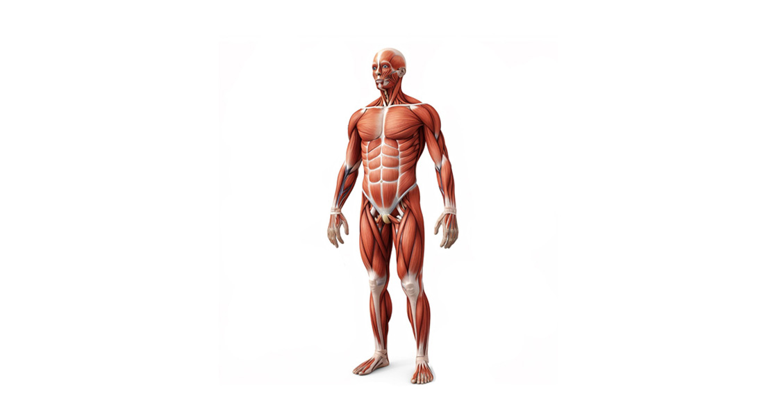
The Muscular System
- The muscular system is an organ system comprised of three types of muscle: skeletal, smooth, and cardiac.
- Functions:
- Permits movement of the body.
- Maintains posture.
- Circulates blood throughout the body.
- The muscular system in vertebrates is primarily controlled by the nervous system, although some muscles function autonomously.
Main Functions of the Muscular System
- Movement:
- Skeletal muscles pull on bones to facilitate movement at joints.
- Support:
- Muscles support the internal organs, particularly in the body wall.
- Protection:
- Muscles help protect vital organs.
- Heat Generation:
- Muscle activity generates heat, helping to maintain body temperature.
- Blood Circulation:
- Cardiac muscles circulate blood through the heart and blood vessels.
Muscle Classification
Muscles are classified based on three criteria:
1. Depending on Striations
- Striated Muscle:
- Contains cross-striations (transverse lines).
- Includes skeletal and cardiac muscles.
- Non-striated Muscle:
- Lacks cross-striations.
- Also known as smooth muscle; found in visceral organs.
2. Depending on Control
- Voluntary Muscle:
- Controlled by conscious will.
- Primarily skeletal muscles; innervated by somatic nerves.
- Involuntary Muscle:
- Cannot be controlled consciously.
- Includes cardiac and smooth muscles; innervated by autonomic nerves.
3. Depending on Situation
- Skeletal Muscle:
- Attached to bones; makes up 40-50% of body mass; voluntary and striated.
- Cardiac Muscle:
- Forms the heart's musculature; involuntary and striated.
- Smooth Muscle:
- Associated with viscera; non-striated; involuntary.
Structure of Muscle
- Muscle Tissue:
- Composed of individual muscle cells, known as myocytes or muscle fibers.
- Muscle fibers are long and slender, multinucleated, and arranged parallel with connective tissue in between.
Connective Tissue Layers
- Fascia: Thick fibrous tissue layer separating muscle mass from neighboring tissues.
- Epimysium: Connective tissue sheath covering the entire muscle.
- Perimysium: Covers bundles of muscle fibers (fasciculi).
- Endomysium: Covers individual muscle fibers.
Muscle Fiber Characteristics
- Cylindrical shape; average length 3 cm (varies between 1-4 cm).
- Diameter ranges from 10 µm to 100 µm.
- Muscle fibers connect to tendons, which attach to bones.
Components of Muscle Fiber
- Nuclei: Multiple nuclei located just beneath the sarcolemma.
- Myofibrils: Fine filaments running through the length of the muscle fiber.
- Mitochondria: Energy production.
- Sarcoplasmic Reticulum: Calcium storage and release.
- Ribosomes: Protein synthesis.
- Glycogen and Lipid Droplets: Energy reserves.
Myofibrils
- Myofibrils are composed of contractile proteins arranged in repeating units called sarcomeres.
- Each myofibril consists of alternating light (I band) and dark (A band) bands.
Sarcomere Structure
- Sarcomere: Basic contractile unit of skeletal muscle, extending between two Z lines.
- I Band: Light band, isotropic to polarized light.
- A Band: Dark band, anisotropic to polarized light.
- Z Line: Protein disk dividing adjacent sarcomeres.
- H Zone: Central light region of the A band, containing only myosin filaments.
- M Line: Center of the H zone, formed by myosin-binding proteins.
Filament Types
- Actin Filaments: Thin filaments extending from Z lines into the A band.
- Myosin Filaments: Thick filaments located in the A band.
Contraction Mechanism
- Cross-Bridges: Projections from myosin filaments that interact with actin.
- Sliding Filament Theory: During contraction:
- Z lines move closer together.
- H zone and I band decrease in size.
- A band remains unchanged.
- Muscle fibers return to their original length during relaxation.

Free Videos